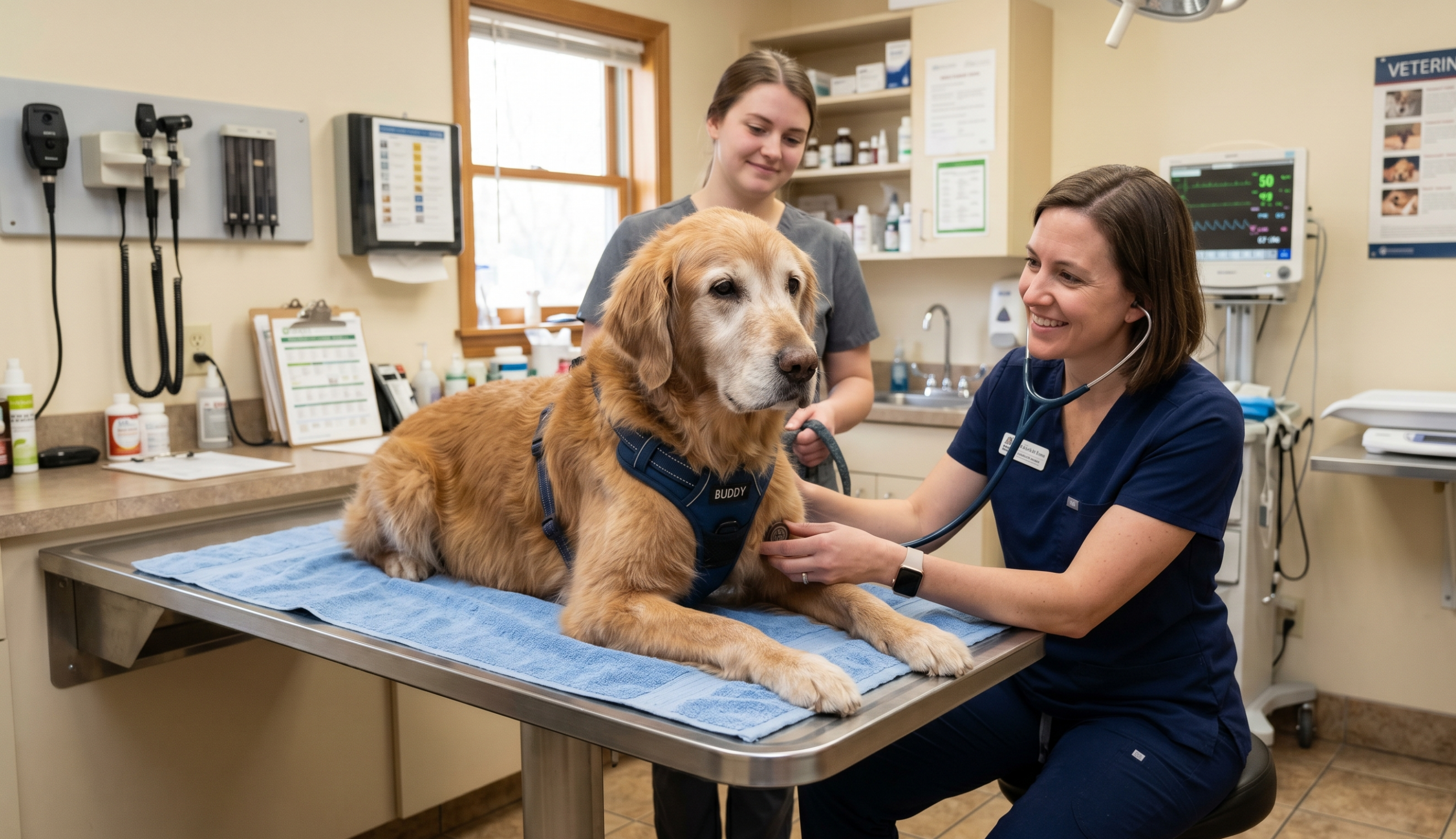

Amniotic membranes for skin wounds in dogs represent a promising area in regenerative veterinary medicine. These membranes provide a biocompatible scaffold rich in bioactive molecules, growth factors, and cytokines, thereby supporting cell growth and proliferation. Their use aims to accelerate wound healing, reduce inflammation, and minimise fibrosis in challenging cases.
Biological Properties and Mechanisms
The amniotic membrane is the innermost fetal membrane, naturally designed to protect and nourish developing tissues. It contains numerous growth factors such as VEGF, bFGF, and cytokines that support angiogenesis, epithelialization, and fibroblast proliferation. These properties make it suitable for managing full-thickness and chronic wounds in veterinary patients.
Clinical Applications and Evidence
Recent studies have demonstrated the efficacy of both fresh and lyophilized amniotic membranes in canine wound healing. In controlled trials, wounds treated with amniotic membranes showed faster closure, reduced exudate, and less pain compared to conventional therapy.
The application of amniotic membrane-derived mesenchymal stem cells or their conditioned media has also led to significant reductions in wound area, particularly in complicated or non-healing wounds.
Practical Considerations and Limitations
Despite their benefits, the widespread clinical adoption of these technologies in veterinary medicine faces challenges. These include sourcing, preservation, and regulatory concerns. Lyophilization helps maintain membrane bioactivity during storage, making clinical use more feasible. Still, more large-scale studies are needed to standardise protocols and confirm long-term safety and efficacy.
Future Directions in Canine Wound Management
As research progresses, amniotic membranes are expected to become more accessible and better integrated into multimodal wound management. Combining these membranes with other regenerative therapies, such as platelet-rich plasma or stem cell products, could further enhance outcomes for canine patients with severe skin wounds.
Step-by-Step Clinical Application Guide for Amniotic Membranes in Canine Wounds
For practitioners considering amniotic membranes, key steps include:
1: Systematic Approach to Wound Management
When incorporating amniotic membranes into wound management for dogs, veterinarians should follow a systematic approach to maximize healing potential.
The first step involves careful patient selection, prioritizing cases such as chronic non-healing wounds, full-thickness injuries with poor granulation, burns, degloving wounds, or surgical defects.
However, wounds with active infection, significant necrotic tissue, or those in systemically compromised patients (e.g., uncontrolled diabetes) should be stabilized before membrane application.
2: Essential Wound Preparation Protocol
Once a suitable case is identified, thorough wound preparation is essential. The wound bed must be cleansed with sterile saline or a dilute antiseptic solution, avoiding harsh agents such as hydrogen peroxide, which can damage healthy tissue.
Debridement, whether surgical, enzymatic, or mechanical, is a procedure that should remove all necrotic material and biofilm to create a clean, vascularized surface. Hemostasis must also be achieved before proceeding to prevent membrane displacement due to bleeding.
3: Selecting the Appropriate Membrane Type
The choice between fresh and lyophilized amniotic membranes depends on availability and the characteristics of the wound. Fresh membranes retain higher concentrations of bioactive factors, making them ideal for acute, highly exudative wounds, but they require immediate use.
Lyophilized membranes, on the other hand, offer extended shelf life and comparable efficacy for chronic wounds, provided they are rehydrated adequately before application.
4: Proper Membrane Application Techniques
During application, the membrane should be trimmed to slightly overlap the wound edges and placed in direct contact with the wound bed to avoid air pockets. Fixation can be achieved using absorbable sutures (4-0 to 5-0), fibrin glue, or veterinary adhesives. A well-padded bandage with a non-adherent primary layer (e.g., silicone mesh) helps secure the membrane while managing exudate.
5: Critical Post-Application Monitoring
Post-application monitoring is critical for success. The first recheck should occur within 48-72 hours to assess membrane adherence and detect early signs of infection. Subsequent evaluations, conducted every 5-7 days, should track granulation tissue formation, epithelial migration, and wound contraction.
Complications such as excessive exudate, membrane displacement, or allergic reactions require prompt intervention. Bandage changes every 2-3 days may be necessary initially, with frequency adjusted based on wound progression.
6: Enhancing Healing with Adjunctive Therapies
To further enhance healing, adjunctive therapies such as negative pressure wound therapy (NPWT), platelet-rich plasma (PRP), or targeted antibiotics (if infection is suspected) can be integrated. Additionally, client education plays a vital role; owners should be instructed on preventing self-trauma (e.g., using Elizabethan collars), providing proper nutrition (high-protein diets with omega-3 fatty acids), and adhering to follow-up schedules.
Key Considerations for Optimal Outcomes
By following these structured steps, veterinarians can effectively incorporate amniotic membranes into wound management protocols, improving outcomes for canine patients with challenging skin injuries.
- Amniotic membranes are not a standalone solution; proper wound preparation and ongoing care are fundamental.
- Lyophilized membranes provide a practical alternative for clinics without access to fresh tissue, with minimal compromise in efficacy.
- Combining amniotic membranes with other regenerative therapies (e.g., PRP, NPWT) may yield superior results in complex cases.
Conclusion: Bridging Research and Practice
Amniotic membranes offer a transformative tool for canine wound care, but their full potential depends on overcoming logistical hurdles and advancing evidence-based protocols. With continued research, they may become a staple in regenerative veterinary medicine.