An evidence-based review of shockwave therapy for canine osteoarthritis, covering mechanisms, device types, protocols, efficacy, and practical clinical guidance.
Canine osteoarthritis (OA) is among the most prevalent and debilitating musculoskeletal conditions in veterinary medicine. Affecting an estimated 20% of adult dogs and up to 80% of geriatric dogs, OA is a leading cause of chronic pain, impaired mobility, and reduced quality of life. Despite the availability of pharmacologic options such as NSAIDs, and nonpharmacologic interventions including weight management and rehabilitation, many dogs continue to experience pain and functional limitations. In recent decades,extracorporeal shockwave therapy (ESWT) has emerged as a promising adjunctive treatment. Don’t let the name scare you – it’s really sound-wave therapy. Originally developed for human medicine in the 1970s for lithotripsy, ESWT was soon explored for musculoskeletal care. By the late 1990s, equine practitioners were using it for tendon, ligament, and bone injuries, and canine applications followed soon after. ESWT is now widely used in human sports medicine, orthopedics, and physical therapy. As a veterinary rehabilitation therapist and pain-management clinician, I value shockwave because it integrates seamlessly — in general practice, specialty settings, and structured rehab programs — and it even works well for mobile practitioners. I often reach for ESWT in end-stage OA or refractory cases that haven’t responded long enough — or well enough — to other modalities I also love, like laser and acupuncture. In my hands, shockwave provides a different “lever” for comfort that complements those tools. My clinical experience is primarily with electrohydraulic focused systems, the same type used in many veterinary studies, but I remain open to new devices as the tech evolves. This article reviews the scientific rationale behind ESWT, along with available technologies, the current evidence base, and practical considerations — including efficacy, cost, sedation requirements, and client decision-making.
Mechanisms of action: why shockwaves work
Shockwaves are focused, high-energy acoustic pressure waves (sound waves) generated outside the body and transmitted through tissues. When the waves encounter tissue interfaces of differing density, such as bone–tendon or cartilage–bone junctions, they release stored energy. The resulting mechanical and biological effects contribute to pain relief and tissue healing.3,6
Biological responses
Research in both experimental models and clinical settings has demonstrated multiple beneficial biological responses to ESWT:
- Neovascularization: Shockwaves stimulate new blood vessel growth, improving tissue oxygenation and nutrient delivery.3
- Cartilage preservation: Reduced nitric oxide production and inhibition of chondrocyte apoptosis help protect cartilage from degenerative changes.
- Bone remodeling: ESWT enhances osteoblastic activity, promoting new bone and cartilage formation, while stimulating osteoclastic activity to remodel bone spurs and mineralized deposits.1
- Analgesic effects: Depletion of substance P and modulation of nociceptors contribute to reduced pain perception.3
- Mechanotransduction: Physical microstress in tissues triggers molecular signaling pathways, releasing cytokines such as TGF-β1 and VEGF and further supporting healing.6
In arthritis specifically, ESWT primarily provides symptomatic pain relief, though it does not halt or reverse disease progression.1
Technology and device types
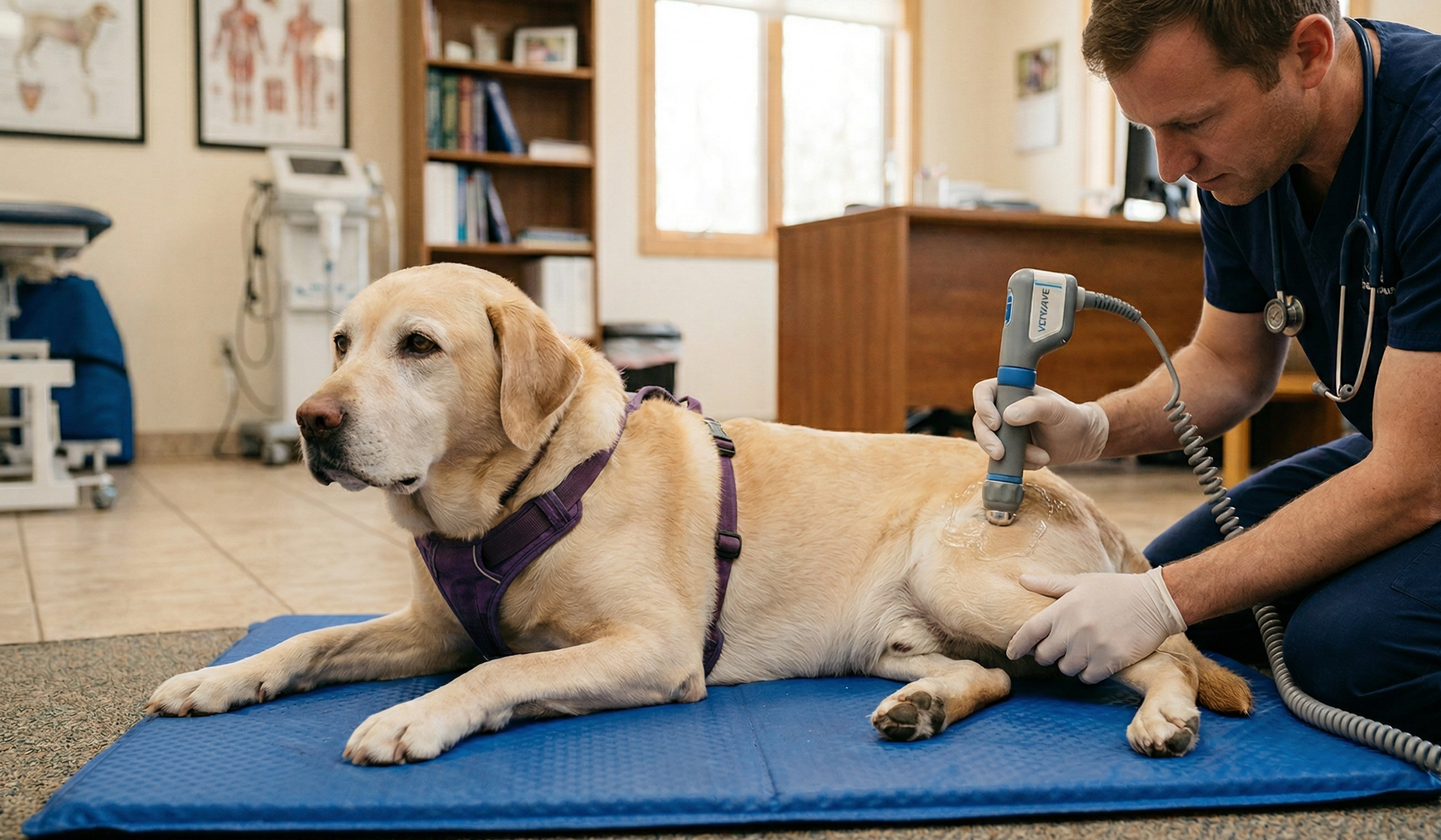
Multiple technologies are used to generate shockwaves, each with distinct engineering mechanisms and tissue penetration profiles: Electrohydraulic ESWT – An electrical spark within a water medium vaporizes molecules, creating an expanding shockwave that is focused via a reflector. This technology was among the first applied in both human and veterinary orthopedics.6 Electromagnetic ESWT – A magnetic coil accelerates a metal membrane, producing a pressure wave that is focused with an acoustic lens. Provides precise, repeatable energy delivery. Piezoelectric ESWT – Spherical arrays of piezoelectric crystals contract and expand under voltage, generating tightly focused pressure waves. Offers pinpoint targeting and less discomfort in surrounding tissue. Radial Pressure Wave Therapy (RPWT) – Unlike focused ESWT, RPWT generates lower-energy radial waves via pneumatic projectile impact. Energy disperses superficially rather than focusing deeply, making RPWT more suitable for tendinopathies or superficial conditions. RPWT is popular in Europe but is not FDA-approved for human musculoskeletal use in the US.1 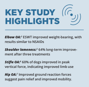 The head of the device is called a trode, and different trode sizes can be chosen depending on the depth of treatment needed. Ultrasound gel is applied on the skin to transmit the sound waves; shaving hair is often unnecessary. Protocols will be reviewed below.
Clinical evidence for shockwave therapy in canine arthritis
Although extracorporeal shockwave therapy (ESWT) has been widely studied in equine and human orthopedics, veterinary literature on canine arthritis is more limited. Nonetheless, several well-designed studies and clinical reports support its role as an effective adjunct therapy for dogs with osteoarthritis (OA).
Elbow osteoarthritis
One of the most rigorous evaluations of ESWT in dogs was conducted at the University of Tennessee.3 In this randomized, controlled trial, dogs with moderate to severe elbow OA were treated with ESWT or sham therapy. All dogs were allowed to remain on stable arthritis medications to avoid confounding effects.
- Protocol: Two ESWT treatments (days 0 and 14), 500 pulses per joint, energy flux density 0.13 mJ/mm.²
- Findings: Dogs receiving ESWT demonstrated significant improvements in weight-bearing measured by force plate analysis, comparable to those expected from NSAID therapy.
- Conclusion: Even in patients already managed medically, ESWT provided additive benefits. The study authors noted the need for further research to refine treatment frequency, energy levels, and protocols.
Shoulder lameness
A retrospective study from Tufts University evaluated ESWT in 15 dogs with shoulder-associated lameness, including instability, calcifying tendinopathies, and inflammatory lesions.2
- Protocol: Three ESWT sessions at three- to four-week intervals, using electrohydraulic devices. Sedation with dexmedetomidine and butorphanol was required for each session.
- Short-term results: Of nine dogs available for final recheck, three had complete resolution of lameness and six showed improvement.
- Long-term follow-up: At an average of 844 days, 64% of owners reported improved or normal function in their dogs. No adverse effects were documented.
- Conclusion: ESWT provided durable improvements in chronic shoulder lameness refractory to conservative management.
Stifle (knee) osteoarthritis
Force plate analysis has been used to evaluate ESWT in dogs with stifle OA. In one study, 60% of treated dogs showed improved peak vertical force compared to baseline, indicating improved limb use.8 Clinical experience suggests stifle responses may be less dramatic than hip or shoulder outcomes, though still beneficial.
Hip osteoarthritis
Dogs with coxofemoral OA treated with ESWT demonstrated improved ground reaction forces compared with baseline, suggesting reduced pain and improved mobility.7 While sample sizes have been limited, results are consistent with findings in equine and human studies.
Overall response rates
Practitioner surveys1 indicate:
- ~70% of dogs respond dramatically to ESWT.
- ~15% show moderate improvement, often enhanced by repeat sessions.
- ~15% show no benefit.
- Shoulders, hips, and backs generally respond best, while stifle (knee) conditions may be less predictable.
In my practice, I consider shockwave when a dog with OA has plateaued on acupuncture, laser, structured exercise, and medication — or when we need a bigger swing at comfort in end-stage joint disease. It’s not a replacement for the basics (managing weight, movement, environment), but it’s a potent complement that often re-opens a window for function.
Safety and adverse effects
ESWT is generally safe when performed according to guidelines. Reported adverse effects are minimal:
- Transient soreness after treatment, considered part of the expected healing cascade.5
- Sedation-related risks, which can be mitigated through safe anesthesia practices such as pre-sedation bloodwork, monitoring vital signs, and providing supportive care during sedation.
- No long-term tissue damage has been reported in canine studies, though high-intensity or prolonged treatments could theoretically injure bone or soft tissue.1
Treatment protocols and practical considerations
ESWT for canine arthritis is typically performed on an outpatient basis, with sedation required for most patients. Protocols vary depending on the device, condition treated, and clinician preference, but some general patterns have emerged:
- Number of sessions: Commonly one to three sessions, spaced two to six weeks apart.2
- Impulses: 500 to 1,500 pulses per treatment, depending on joint size.3
- Energy Flux Density (EFD): Generally between 0.1 and 0.3 mJ/mm² for canine arthritis studies.3
- Duration: Treatments generally last five to 15 minutes per joint, with newer units capable of even shorter procedures.
- Treatment course: Noticeable improvements are often reported within one to two weeks after the first treatment, and cumulative benefits continue following successive sessions.
Most manufacturers provide suggested treatment settings for common indications, though optimization of veterinary-specific protocols remains an area of active research.
Sedation and patient management
Earlier generations of electrohydraulic ESWT units produced loud percussive sounds and high peak pressures, which often required sedation or anesthesia for safe, accurate, and humane treatment. Common veterinary sedation protocols included dexmedetomidine (5–10 µg/kg) and butorphanol (0.2 mg/kg).2 Important considerations for sedation:
- Patient safety: Sedation adds a layer of risk, particularly in geriatric dogs with comorbidities.
- Time commitment: Owners should anticipate a procedure-day commitment, similar to radiographs or dentistry.
- Recovery: Most dogs recover uneventfully and can return home the same day.
With advances in technology — particularly acoustic dampening, controlled pulse shaping, and broader focal areas — modern ESWT can now be performed in awake, cooperative dogs under specific circumstances, particularly with systems engineered for comfort and energy uniformity. As a Fear Free practitioner, my clinic is designed to be as peaceful as possible, with quiet music playing to calm patients and mask outside noise pollution. With that in mind, it’s no surprise that shockwave isn’t my favorite from a sound-environment standpoint — but I’m also a sound-therapy practitioner and see tremendous value in this acoustic-based modality when chosen thoughtfully. I’ll treat patients awake only if they are relaxed and tolerant; otherwise, I recommend sedation to prevent fear or struggling. I prefer to avoid sedation in senior patients with renal or cardiac disease — in those cases we proceed only if the dog is truly comfortable awake, or pivot to other modalities.
Cost considerations
Cost is one of the most significant barriers to widespread use of ESWT.
- Equipment investment: True shockwave units range from $20,000 to $30,000 or more, and some require ongoing maintenance which can also be costly. This makes acquisition challenging for smaller practices.
- Lack of insurance reimbursement: Unlike human medicine, ESWT for veterinary patients is generally out-of-pocket for owners.
- Client cost per session: Fees vary by region and practice but commonly range from $300 to $500 per treatment. With two to three treatments recommended, the total client investment can exceed $1,000.
Some clinics offset costs by combining ESWT with other rehabilitation services (laser therapy, underwater treadmill) in bundled packages, improving owner-perceived value. 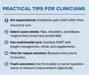
Duration of effect and re-treatment
- Onset of relief: Many dogs show improvement within one to two weeks.
- Duration of effect: Pain relief typically lasts six to 12 months; some patients may need annual or semi-annual re-treatment.
- Repeatability: ESWT can be safely repeated when clinical signs return, making it a sustainable option for long-term management.
Client communication
Because ESWT is relatively new to many pet owners, effective communication is essential. Owners should understand that:
- ESWT is adjunctive, not curative. It reduces pain but does not stop OA progression.
- Most dogs respond well, but a minority may show little or no improvement.
- Sedation may be needed, but risks are low when appropriately managed.
- Costs are significant, but relief can be long-lasting and reduce reliance on daily medications.
Pros and cons summary
Advantages
- Noninvasive, outpatient treatment
- Durable pain relief that often lasts months
- May reduce reliance on daily NSAIDs and other systemic meds
- Generally safe with minimal adverse effects reported in studies
- Integrates well with multimodal OA care (rehab, acupuncture, laser, weight management)
Disadvantages
- May require sedation or anesthesia in anxious or multi-joint case
- High equipment cost; treatments can be expensive for client
- Maintenance can add up: some systems require trode replacement (e.g. after ~50,000 shocks, depending on model
- Variable response rate: ~15% of dogs may show little or no benefi
- Often not covered by pet insurance (owner pays out-of-pocket
- Noise can be stressful if sedation is inadequate or the patient is not mellow.
Electrohydraulic vs. piezoelectric systems
Current veterinary ESWT technologies primarily use either electrohydraulic (EH) or piezoelectric (PE) mechanisms. Both generate therapeutic shockwaves, but each has distinct performance characteristics.
| Feature | Electrohydraulic | Piezoelectric |
| Mechanism | Spark discharge in water reflected into focal zone | Crystalline compression generating precise acoustic pulse |
| Focal area | Broad treatment field, ideal for joints and bony interfaces | Small, sharply defined zone for precision targeting |
| Penetration depth | Deep, effective for hips, elbows, spine | Shallower, optimal for tendons and focal lesions |
| Sedation requirement | Optional with modern dampening | Optional due to low surface discomfort |
| Consistency | Minor variation pulse-to-pulse; electrodes wear over time | Consistent pulse delivery across lifespan |
| Maintenance | Electrodes last ~20,000–50,000 shocks; replacement ~$2,000 | No electrode wear; higher initial unit cost |
| Cost per use | Lower per-pulse for high-volume practices | Higher upfront cost but lower maintenance |
| Applications | Best for large joints and orthopedic disease | Best for tendinopathies and localized lesions |
- Electrohydraulic (EH) systems remain the gold standard for orthopedic applications. The broad focal field enables effective treatment of large joints and bony structures.Theelectrohydraulic source generates uniform, high-energy waves while minimizing discomfort through acoustic dampening and pulse shaping.
- Piezoelectric (PE) systemsuse piezoelectric crystals to deliver highly consistent, precisely focused pulses. They require minimal maintenance and rarely need sedation but cover smaller focal zones. PE technology offers outstanding precision for localized tendon or ligament pathology, though it’s less efficient for diffuse joint disease.
My clinical experience is mostly with the electrohydraulic focused systems used in the majority of published canine studies. For deep, broad joint diseaselike end-stage OA, I find EH’s larger focal zone efficient. For very focal soft-tissue problems, a piezoelectric profile can be attractive. I’m brand-agnostic long-term: as protocols and devices evolve, I’ll use what best serves the patient and my practice.
Post-treatment care
Transient tenderness may occur for 24 to 48 hours post-procedure. Dogs should avoid strenuous activity for several days. Clinical improvement is often evident within a week, with progressive gains over four to six weeks as inflammation subsides and tissue remodeling begins.
Practical recommendations for clinicians
- Select technology by indication: Use electrohydraulic systems for arthritis and deep orthopedic lesions; piezoelectric systems for tendon/ligament disorders.
- Assess patient temperament: Sedation-free therapy is achievable in many cases with modern EH systems.
- Combine modalities: Incorporate ESWT into multimodal OA management with acupuncture, laser therapy, and physical rehabilitation.
- Educate clients: ESWT relieves pain and may slow disease progression, but it does not reverse OA.
- Document outcomes: Use force-plate analysis, range-of-motion testing, or owner-reported pain scores to quantify improvement.
Technological advances and future outlook
Recent advances — such as integrated imaging, improved acoustic control, and ergonomic applicators — have enhanced both therapeutic precision and patient comfort.5,6 Systems that combine deep penetration and sedation-free operation mark a new era in companion-animal orthopedics — bridging high efficacy with patient-centered comfort. As veterinary research continues, standardized dosing and comparative trials between EH and PE systems will further refine shockwave therapy for canine osteoarthritis and shockwave’s role in multimodal osteoarthritis management.
Conclusion
Shockwave therapy for canine osteoarthritis has emerged as a valuable adjunct in the management of the condition. By harnessing high-energy acoustic waves, ESWT reduces pain and enhances mobility through a combination of biological effects — including neovascularization, modulation of inflammation, and stimulation of bone and cartilage remodeling.2-4 Clinical studies demonstrate improvements in weight-bearing in elbow OA, long-term function in shoulder lameness, and enhanced mobility in hip and stifle arthritis. Outcomes of shockwave therapy for canine osteoarthritis are often comparable to those achieved with NSAIDs, yet without the risks of long-term drug use. 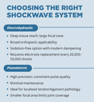 Despite its promise, ESWT is not without limitations. Equipment costs remain high, client fees significant, and sedation is often required. Moreover, response rates of shockwave therapy for canine osteoarthritis vary, and standardized protocols are still evolving. Nonetheless, with reported benefits lasting six to 12 months, repeatable treatments, and minimal side effects, ESWT represents a compelling option within a multimodal arthritis management plan. As veterinary technology continues to advance, not only shockwave therapy shockwave therapy for canine osteoarthritisy will likely become more accessible and refined. For now, it offers hope for dogs whose pain persists despite conventional therapies, and it provides veterinarians with a powerful tool to improve patient comfort and quality of life.
References
1Straus M. Shock wave therapy: Shocking news for dogs with arthritis. Whole Dog Journal. 2004. 2Becker W, Kowaleski MP, McCarthy RJ, Blake CA. Extracorporeal shockwave therapy for shoulder lameness in dogs. J Am Anim Hosp Assoc. 2015;51(1):1-7. doi:10.5326/JAAHA-MS-6175. 3Millis DL. Extracorporeal shockwave therapy for elbow DJD. Proceedings, Univ. Tennessee College of Veterinary Medicine; 2005. 4Kirkby K. Shockwave therapy as a treatment option. Clinician’s Brief. August 2013:51-55. 5Marovino T. Top 10 must-have devices for your practice: Extracorporeal shockwave therapy. Practical Pain Management. October 2014:29-32. 6Ioppolo F, Rompe JD, Furia JP, Cacchio A. Clinical application of shock wave therapy in musculoskeletal disorders. Eur J Phys Rehabil Med. 2014;50(2):217-230. 7Mueller M, Bockstahler B, Skalicky M, Mlacnik E, Lorinson D. Effects of radial shockwave therapy on the limb function of dogs with hip osteoarthritis. Vet Rec. 2007;160(22):762-765. 8Dahlberg J, Fitch G, Evans RB, McClure SR, Conzemius M. The evaluation of extracorporeal shockwave therapy in naturally occurring osteoarthritis of the stifle joint in dogs. Vet Comp Orthop Traumatol. 2005;18(3):147-152