
Findings from a recent study on splenic lesions may improve diagnostic management of splenic neoplasia.
The spleen, often overlooked, but vitally important, can be the site of various lesions in dogs. These lesions can be neoplastic or non-neoplastic in nature, and manifest in different forms, including diffuse, nodular, or multinodular growth, posing diagnostic challenges for veterinarians. New research has investigated the microscopic diagnosis of splenic lesions, aiming to establish correlations between tumor size, number of nodules, and specific diagnoses, thereby enhancing our understanding and diagnostic approach to these conditions.
Study Involved Characterization of Splenic Masses
In a recent study, researchers sought to characterize splenic masses in dogs and identify potential risk factors associated with splenic tumors. One of the studys key findings was the prevalence of hemangiosarcoma as the most common neoplastic diagnosis, while hyperplasia emerged as the predominant non-neoplastic condition. These findings provide valuable guidance for veterinarians navigating splenic lesions in canine patients.
Demographic Profile Suggests Which Dogs Most Affected
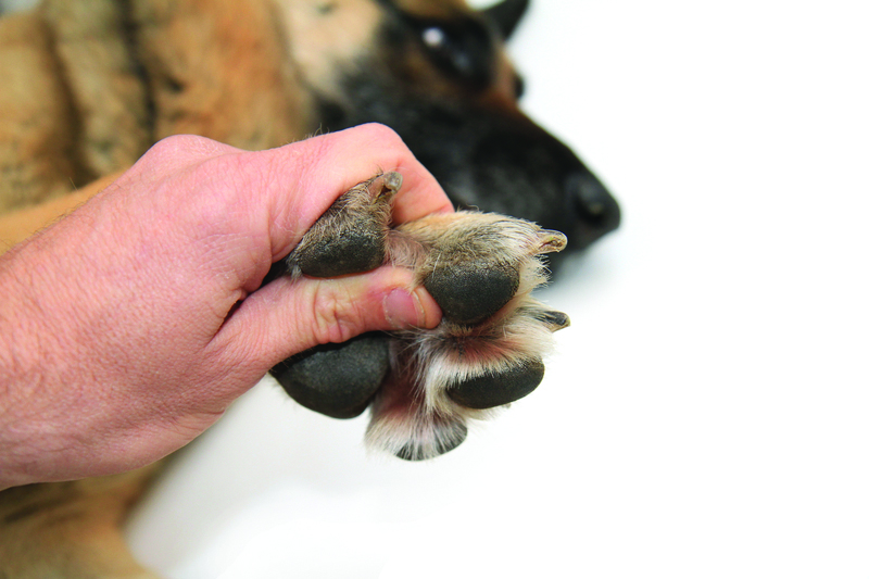
Understanding the demographic profile of affected dogs can also aid in early detection and management. The study revealed that male, senior, and purebred dogs, including breeds such as the Cocker Spaniel, German Shepherd, and Labrador Retriever, were more commonly affected. This demographic predisposition sheds light on the importance of targeted screening and surveillance in these populations, potentially leading to earlier intervention and improved outcomes.
Additional Risk Factors
The study also identified specific risk factors associated with the development of splenic neoplasia. Male sex was found to be a significant risk factor, emphasizing the need for heightened vigilance and proactive screening in male dogs. As well, the presence of two or more splenic nodules, and an increase in nodule size greater than 2 cm, were identified as indicating increased risk for splenic neoplasia. Recognizing these factors enables veterinarians to prioritize diagnostic investigations and tailor treatment strategies accordingly.
Importantly, these findings highlight the value of comprehensive diagnostic approaches in cases of splenic lesions. While imaging techniques play a crucial role in detection, histopathological examination remains the gold standard for accurate diagnosis. By integrating clinical, imaging, and histopathological data, veterinarians can formulate precise diagnostic and therapeutic plans, optimizing patient care and outcomes.
This study offers valuable insights into the characterization and risk factors associated with splenic lesions in dogs. Veterinarians can enhance their diagnostic acumen and therapeutic interventions by leveraging this knowledge, ultimately improving the management of canine patients with splenic abnormalities. Continued research in this field promises to further refine our understanding and approach to these complex conditions, benefiting both veterinary practitioners and their canine patients.