
The study’s emphasis on atypical mitotic figures introduces a microscopic parameter that holds promise in enhancing the precision of survival predictions.
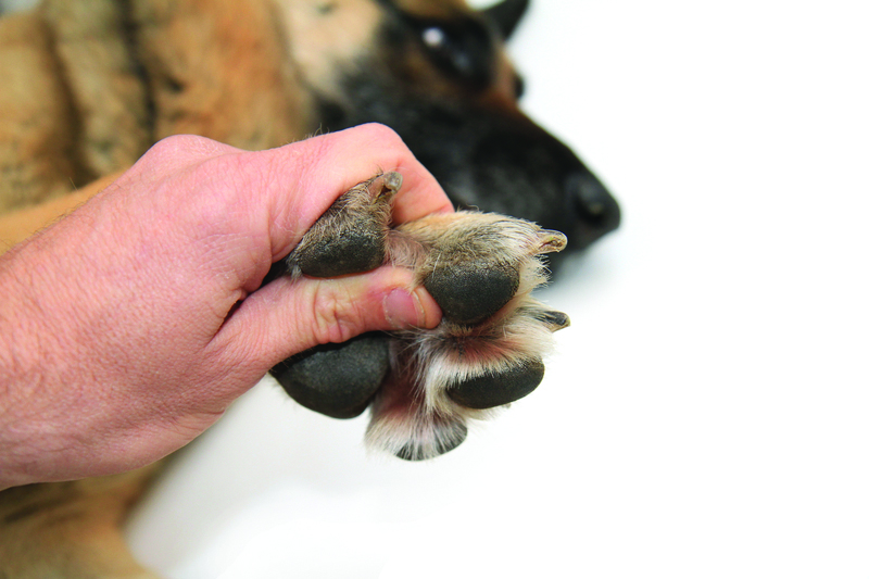
Mast cell tumors stand out as one of the most prevalent skin tumors in canines. Understanding the prognosis for these tumors is crucial for effective management, and a recent study has introduced a novel microscopic parameter atypical mitotic figures to assess survival predictions.
Mitotic figures, the counting of dividing tumor cells, have long been a key factor in determining the prognosis of mast cell tumors in dogs. However, this study introduces a unique approach by specifically examining atypical mitotic figures, shedding new light on their prognostic significance.
Background: Mast Cell Tumors in Dogs
Mast cell tumors, arising from mast cells in the skin, are a common occurrence in dogs. Mitotic figures, which represent cells undergoing division, have traditionally been scrutinized to gauge the aggressiveness of these tumors. The study, aiming to refine this evaluation, delves into the realm of atypical mitotic figures.
Atypical Mitotic Figures: Unraveling the Microscopic Criterion
Unlike normal cells, tumor cells may exhibit errors during chromosome separation, leading to atypical mitotic figures. These figures serve as a microscopic criterion, often associated with malignancy in various human tumors. The study’s innovation lies in applying this concept to canine cutaneous mast cell tumors.
Key Findings and Prognostic Insights
The research demonstrates a direct correlation between a high number of atypical mitotic figures and shorter patient survival in canine cutaneous mast cell tumors. Unlike traditional mitotic figure counting, the presence of increased atypical mitotic figures (?3 per 2.37 mm2) proves to have higher specificity for predicting tumor-related death.
Future Implications and Validation
While this study marks a significant stride in prognostic evaluation, the findings call for further validation in subsequent studies. The potential for atypical mitotic figures to refine prognostic assessments in canine mast cell tumors opens avenues for more targeted and accurate predictions, guiding veterinarians in devising tailored treatment plans.