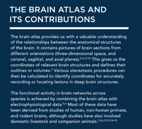
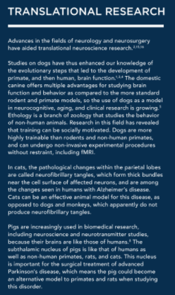
Combining data from the brain atlas and MRI/fMRI neuroimaging can help shed light about the brains of companion animals, horses, and pigs. ?
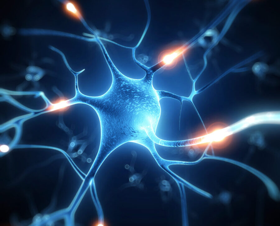
When it comes to understanding animal brains and their structural, physiological, and behavioral functions neuroimaging techniques such as magnetic resonance imaging (MRI) and functional MRI (fMRI) play a major role.1,2,3,4 These advances help both veterinary researchers and clinicians understand how different areas of the brain communicate and integrate.7 ?
This article combines data from the brain atlas and MRI/fMRI neuroimaging of companion animal species, horses, and pigs,3,4,6,7-9 for understanding animal brains, and the differences and similarities between them.??
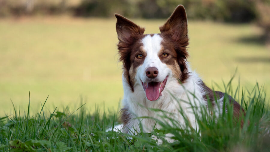
Exploring the brains of dogs?
The canine brain is gyrencephalic (having cortical folds), making it more like the human brain than rodent and avian models, which are lissencephalic (without cortical folds).5 Lets take a closer look at what weve learned about the canine brain:
1) The role of the cingulate cortex and lateral frontal lobes
Resting (task-free) fMRI investigations of brain activity have identified the important role of the cingulate cortex in the evolution of the mammalian brain.??
The cingulate cortex is a bilateral structure located deep within the brains cerebral cortex. Over many centuries, its central role and function have been partly taken over by the lateral frontal lobes, which control problem-solving, task-switching, and include word detection and odor distinction. Therefore, brain areas function in synchrony to form task-specific networks. It is interesting to note that these networks have a smaller role in dogs than in humans, despite the proportionately larger canine brain.?
The use of fMRI can help analyze diseases characterized by dysfunctional integration and communication between brain areas. It also can determine which areas of the canine brain are active when the dog reacts to external stimuli.?
2) The functional brain
Dogs are strongly motivated to learn tasks by mimicking the work of another already trained dog because of the praise they receive.3,4 This is evident during testing. During MRI or fMRI, they can lie still without moving for eight minutes when expecting the petting and treats observed with other dogs. Sounds also are played to study which brain areas are activated by various sounds. But functional brain activities are irregular and may not follow any anatomical regular boundaries. Parts of the brain act in synchrony to form a functional brain network.4?
3) The connection between human and canine brains
Aging studies in dogs, like those in humans, have revealed increased framewise displacement (i.e. head motion) within the MRI scanner.3,4 A recent task-free large study involved 33 family trained dogs (1 to 14 years of age, 17 females, 16 males, and various breeds including nine Border Collies, seven Golden Retrievers, five mongrels, two Australian Shepherds, and one each of several other breeds).? The older dogs were somewhat less able to maintain their initial position as shown by head displacement. Similar findings are seen in older people, as normal aging results in increased head motion.4???
These family dogs were studied as a population group not bred or reared under laboratory research and testing conditions. They lived in their natural social environments, which provided useful data for awake fMRI assessments.?
The effects of human intervention on the canine brain
Significant neuroanatomical variation has been documented among a variety of dog breeds based on how they were bred i.e. for specific tasks such as hunting, herding, guarding, or companionship10 Has this purpose-bred approach altered the gross anatomical organization of the canine brain??
The reported variation in the study was distributed non-randomly across the brain and was plainly visible on stereotactic analysis. Specific regional sub-networks co-varied significantly, and not merely because of variations in total brain or body size, or skull shape. The behavioral specialization selected for within the four functional dog groups (hunting, herding, guarding, companionship) significantly correlated with their anatomy, and most changes occurred in the terminal branches of the phylogenetic tree, indicating a strong recent selection in these individual breeds. Thus, through selective breeding, humans have differentially altered the brains of various lines of domestic dogs.10???
Interestingly, the authors indicated that based on their published findings, studies involving domesticated animals should not lump them together as a species but should consider breed types, structure, and function.??
What research tells us about feline brains
As brain atrophy increases with aging, studies of older cats documented behavioral changes with more cognitive dysfunction.1 The changes in behavior include excessive vocalization, spatial and/or temporal disorientation, alterations in interactions with owners or other pets, shifts in the sleep-wake cycle, house soiling, changed activity levels, anxiety, and/or learning and memory deficits. Other concurrent disorders include hypertension, hyperthyroidism, pain, and separation anxiety. Atrophy of regional brain structures such as the hippocampus can be viewed with MRI, but the assessment of global atrophy remains difficult, especially because cats are often very stressed during veterinary clinic visits.?
In humans, global and regional brain abnormalities can be assessed with Voxelbased morphometry (VBM).? A study looking at the use of this technology in cats without neurological or behavioral signs (65 cats, aged 17 to 200 months) showed a significant decrease with aging in the gray matter within the brains bilateral parietal lobes. The white matter did not show any size reduction, nor was there a significant age-related reduction in inter-thalamic adhesion.1??
Aging feline brains have shown neuronal loss and atrophy, amyloid-? deposition, and tau phosphorylation. Amyloid-? deposition and tau phosphorylation have been seen not only in the brains parietal cortex, but also in the para-hippocampal cortex and occipital cortex.1?
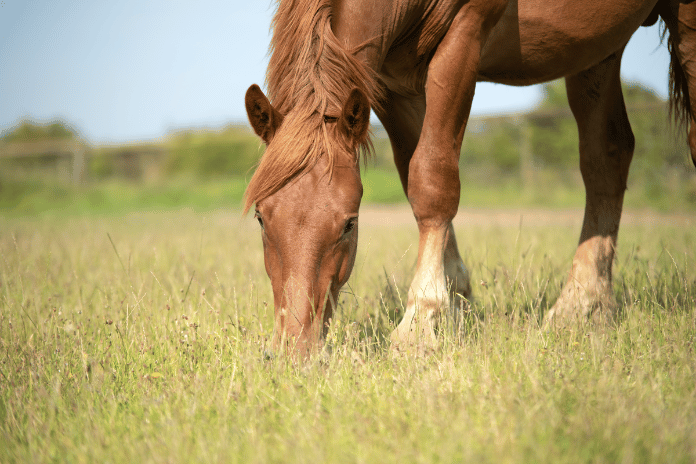
The use of neuroimaging in horses?
Large animal veterinary neuroimaging has become increasingly important, especially in the horse.11 Intracranial diseases in horses with neurological signs are now more often studied by MRI.11,12 High magnetic field strengths (3Tesla) are now available in veterinary medicine; images collected with 3Tesla give improved image clarity and detail. However, even with progress in the technical adaptations of scanners and detection coils used in equine medicine, the description of brain morphology in horses has lagged behind that of other species.1 Completed studies have mostly examined the brains of foals.?
Ungulate animal brain
The characteristics of the ungulate (hoofed animal) brain include a readily seen expanded neocortex and intensive gyrification by MRI. Equine gyri and sulci display a more complex pattern than in carnivores and other ungulates, a finding associated with their increased body mass, and special senses. Sensory information conveyed from the lips is important, and is processed in the cortex surrounding the diagonal gyrus. The nostrils and tongue are represented in the area behind the supra-sylvian sulcus.11,12 The temporal lobe in horses is expanded in such a way that the insular cortex is deeply hidden in the depths of the sylvian and oblique sulcus, and thus is no longer visible from the outer surface.?
The horse is a macrosmatic mammal (highly developed) when it comes to olfactory function. They have a relatively large rhinencephalon and rather small olfactory bulbs. In other ungulates, like the pig and cow, the linkage between these two structures is stronger.11,12?
The hippocampus and para-hippocampal gyrus also appear small in relation to the very large rhinencephalon (olfactory brain). The horses comparatively underdeveloped efferent system, which carries motor signals away from the brain to the rest of the body, could explain the animals minimal motor control of the distal limbs. A characteristic feature of the equine cerebellum is the deviation of the vermis, which controls body posture and locomotion, although the cerebellar shape is characteristic of that seen in ungulates. Some of these published differences may be artefacts.12?
The anatomy of pig brains is similar to humans
The digitalized brain atlas, along with MRI and fMRI, and histological resources have contributed to recent porcine research. The pig brain is large, with cerebral structures common to other mammals, and appears comparable to the human brain in its anatomy, histology, myelination, growth and development, and vascularization (e.g. humans and pigs share a similar cerebrovascular anatomy of the circle of Willis).8 Further, the pig brain more closely resembles that of primates rather than rats in terms of cortical convolutions, shape and number of neocortical neurons.?
Porcine folded brain cortical surface
The pig brain, like that of the human and dog, is gyrencephalic with a folded brain cortical surface and well-defined circumvolutions, while the rat and avian brain are lissencephalic with a smooth-surfaced cerebral cortex.8 The primate brain has a more pronounced curvature of the telencephalon and more developed anterior pole, although the pig brain has a more developed olfactory system that fills a large portion of the anterior brain. The large rostral snout of the pig likely developed for its tactile ability to explore the environment (think searching for truffles).8?
Conclusion
Understanding animal brains of various species through neuroimaging and data related to the brain atlas reveals interesting differences and similarities, and will hopefully lead to beneficial outcomes in the future.
1 Hamamoto Y, Yu Y, Asada R, Mizuno S, Hasegawa D. Age-related brain atrophy in cats without apparent neurological and behavioral signs using voxel-based morphometry. Front. Vet. Sci. 2022, 9:1071002. doi: 10.3389/fvets.2022.1071002.??
2 Radhakrishnan H, Ubele MF, Krumholz SM, Boaz K, Mefford JL, Jones ED, Meacham B, Smiley J, et al. Tacrolimus protects against age-associated microstructural changes in the beagle brain. J. Neurosci. 2021, 41(23):51245133. doi.org/10.1523/JNEUROSCI.0361-21.2021.??
3 Saccà V, Sarica A, Quattrone A, Rocca F, Quattrone A, Novellino F. Aging effect on head motion: a machine learning study on resting state fMRI data. J. Neurosci. Methods 2021, 15;352:109084. doi:10.1016/j.jneumeth.2021.109084 ? Epub 2021 Jan 25. PMID: 33508406.?
4 Szabó D, Janosov M, Czeibert K, Gácsi M, Kubinyi E. Central nodes of canine functional brain networks are concentrated in the cingulate gyrus. Brain Struct. Funct. 2023, 228:831843. doi.org/10.1007/s00429-023-02625-y.?
5 Johnson PJ, Luh W-M, Rivard BC, Graham KL, White A, FitzMaurice M, Loftus JP, Barry EF. Stereotactic cortical atlas of the domestic canine brain. Sci. Rep. 2020, 10:4781. doi.org/10.1038/s41598-020-61665-0.?
6 Banstola A, Reynolds JNJ. Mapping sheep to human brain: The need for a sheep brain atlas. Front. Vet. Sci. 2022, 9:961413. doi: 10.3389/fvets.2022.961413.?
7 Ella A, Barrière DA, Adriaensen H, Palmer DN, Melzer TR, et al. The development of brain magnetic resonance approaches in large animal models for preclinical research. Animal Front. 2019, 9(3): 44-51. doi: 10.1093/af/vfz024.?
8 Sauleau P, Lapouble E, Val-Laillet D, Malbert C-H.? The pig model in brain imaging and neurosurgery. Animal 2009, 3(8):11381151. doi:10.1017/S1751731109004649.?
9 Buxton DF, Compton RW (1986).The canine brain: Basic atlas for an autotutorial approach to the central nervous system. S.I. 34.?
10 Hecht EE, Smaers JB, Dunn WD, Kent M, Preuss TM, Gutman DA. Significant neuroanatomical variation among domestic dog breeds. J. Neurosci. 2019, 39(39):7748-7758. doi.org/10.1523/JNEUROSCI.0303-19.2019.?
11 Schmidt MJ, Knemeyer C, Heinsen H. Neuroanatomy of the equine brain as revealed by high-field (3Tesla) magneticresonance-imaging. PLoS ONE 2019, 14(4): e0213814. doi.org/10.1371/journal.pone.0213814.?
12 Arencibia A, Vazquez JM, Ramirez JA, Ramirez G, Vilar JM, Rivero MA, Alayon S, Gil F. Magnetic resonance imaging of the normal equine brain. Vet. Radiol. 2001, 42(5):405-408. doi.org/10.1111/j.1740-8261.2001.tb00959.x.?
13 Kraft SL, Gavin PR, Wendling LR,?Reddy VK. Canine brain anatomy on magnetic resonance images. Vet. Radiol. 1989, 30(4): 1740-8261. doi.org/10.1111/j.1740-8261.1989.tb00767.x.?
14 Datta R, Lee J, Duda J, Avants BB, Vite CH, et al. A digital atlas of the dog brain. PLoS ONE 2012, 7(12): e52140. doi:10.1371/journal.pone.0052140.?
15 Hudson LC,?Cauzinille? L, Kornegay JN,?Tompkins MB. Magnetic resonance imaging of the normal feline brain. Vet. Radiol. 1995, 36(4):267-275. doi.org/10.1111/j.1740-8261.1995.tb00261.x?
16 Salazar I. The brain of the dog in section: a comprehensive view for veterinary students. Anatomia, Histologia, Embryologia 2005, 34(S1):44.? doi.org/10.1111/j.1439-0264.2005.00669-100.x?
17 De Sousa AA, Dames BAR, Graff EC, Mohamedelhassan R, Vassilopoulos T, Charvet CJ. Going beyond established model systems of Alzheimers disease: companion animals provide novel insights into the neurobiology of aging. Comm. Biol. Perspective 2023, 6 (655): 1-16. doi.org/10.1038/s42003-023-05034-3?
18 Tannenbaum J, Bennett BT. Russell and Burch’s 3Rs then and now: the need for clarity in definition and purpose. J. Am. Assoc. Lab. Anim. Sci. 2015, 54(2):120-132. PMID: 25836957; PMCID: PMC4382615.?