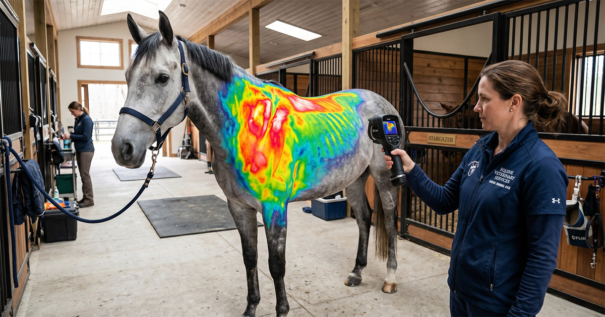
Modern equine wellness is evolving quickly, and one technology gaining attention among horse owners, trainers, and veterinarians is thermal imaging. Once used primarily in industrial and military settings, thermal imaging cameras are now helping equestrians detect subtle changes in a horse’s body before they become obvious injuries or performance problems.
By identifying temperature variations linked to inflammation, circulation, and muscle stress, thermal imaging offers a non-invasive way to support preventative care and long-term soundness.
Detecting Inflammation Early
Inflammation often develops before visible swelling or lameness appears. Thermal imaging works by detecting heat patterns emitted from the horse’s body, allowing caretakers to spot asymmetries or abnormal hot spots that may indicate strain, injury, or irritation.
For example, increased heat in a tendon, hoof, or joint may suggest:
- Early soft tissue stress
- Developing hoof abscesses
- Joint inflammation
- Pressure-related soreness
- Compensatory movement patterns
Because horses naturally mask discomfort, catching these changes early can help owners intervene before a minor issue becomes a major setback.
Thermal scans are especially useful after intense training, competitions, or changes in workload when the risk of strain is higher.
Saddle Fit Applications
One of the most practical uses of thermal imaging in equine care is saddle fit assessment. Poor saddle fit can create uneven pressure points that restrict movement, reduce performance, and contribute to chronic soreness.
After riding, thermal imaging can reveal areas of excessive heat caused by friction or pressure beneath the saddle. Uneven heat patterns may indicate:
- Bridging
- Pinching at the withers
- Pressure along the back muscles
- Imbalanced rider weight distribution
- Poor flocking or saddle structure
When combined with professional saddle fitting, thermal imaging provides an additional layer of information that may help improve comfort and reduce musculoskeletal stress.
Many bodyworkers and saddle fitters now use thermal cameras as part of a broader evaluation process rather than relying only on visual inspection.
Lameness Monitoring
Thermal imaging is increasingly being used to monitor horses recovering from injuries or managing chronic conditions. Since inflammation often produces increased surface temperature, repeated scans can help track healing progress over time.
Thermography may assist in monitoring:
- Tendon and ligament recovery
- Hoof imbalances
- Back soreness
- Arthritic changes
- Post-exercise muscle recovery
Some veterinarians also use comparative imaging between limbs to identify subtle temperature differences that may correspond with uneven loading or developing lameness.
While thermal imaging cannot diagnose lameness on its own, it may help identify areas that require further examination.
Veterinary Integration
Thermal imaging works best when integrated into a comprehensive veterinary and wellness program. It should be viewed as a supportive assessment tool rather than a standalone diagnostic method.
Veterinarians may combine thermography with:
- Physical examinations
- Flexion tests
- Ultrasound imaging
- Digital radiographs
- Gait analysis
- Bloodwork and metabolic testing
Because thermal imaging is non-contact and stress-free, it can also be useful for sensitive horses that become anxious during traditional evaluations.
In performance barns, routine thermal scans are sometimes used proactively to establish baseline patterns for each horse. Monitoring changes over time may help detect subtle issues earlier than observation alone.
Limitations of Thermal Imaging
Despite its growing popularity, thermal imaging has important limitations. Surface temperature changes do not always indicate injury, and many external factors can influence results.
Variables that may affect scans include:
- Ambient temperature
- Sun exposure
- Sweat and moisture
- Dirt or mud
- Clipped versus unclipped coats
- Recent exercise
- Circulation differences
Thermal imaging also cannot determine the exact cause of inflammation. A hot spot may reflect anything from muscle soreness to infection, nerve irritation, or environmental heat exposure.
For this reason, thermography should never replace veterinary diagnosis or appropriate medical imaging. Instead, it functions best as an early warning system that encourages further investigation when needed.
Final Thoughts
Thermal imaging is becoming an increasingly valuable tool in modern equine care, particularly for early detection, saddle fit evaluation, and performance monitoring. Its non-invasive nature makes it appealing for preventative wellness programs focused on keeping horses comfortable and sound.
While it is not a cure-all or diagnostic replacement, thermal imaging can provide meaningful insights when interpreted by trained professionals and combined with traditional veterinary care. As technology becomes more accessible, thermography may continue shaping how horse owners approach proactive health management.