Diaphragmatic hernias are relatively uncommon in cats, and their diagnosis can pose a challenge due to their varied presentations. This blog post presents a case study of an 18-month-old neutered male domestic shorthair cat with a unique type of diaphragmatic hernia known as a “true diaphragmatic hernia” or “pleuroperitoneal hernia.” We explore the cats diagnostic journey, surgical intervention, and the significance of peritoneography in identifying and differentiating diaphragmatic hernias.
Unusual Presentation and Diagnostic Clues
The cat presented after falling from the second floor and sustaining minor traumatic injuries. Routine radiographic examination raised suspicion of a diaphragmatic hernia, but the appearance of circumscribed soft tissues in the thorax was atypical for a classic traumatic diaphragmatic hernia. Further investigation was required to confirm the diagnosis and determine the nature of the hernia.
The Role of Positive Contrast Peritoneography
Positive contrast peritoneography, a technique involving the injection of contrast material into the peritoneal cavity, was instrumental in highlighting the presence of a hernial sac in the cat. The peritoneogram strongly suggested the presence of a true diaphragmatic hernia. This technique played a vital role in differentiating between an acquired traumatic diaphragmatic hernia and a congenital form.
Confirmation and Surgical Intervention
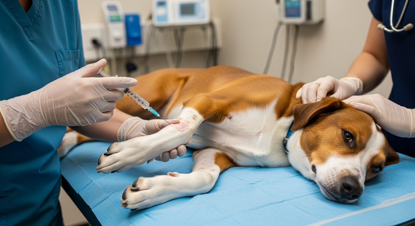
During laparotomy, a 3cm radial diaphragmatic defect was visualized in the right ventral quadrant of the pars sternalis. The edges of the diaphragm were rounded, and a portion of the falciform ligament and omentum were protruding through the defect, contained within a hernial sac. Herniorrhaphy, the surgical repair of the hernia, was successfully performed, and the cat recovered without complications.
Analogous to Morgagni Hernia in Humans
The location of the diaphragmatic defect, ventrally and to the right, is analogous to Morgagni hernia observed in humans. While only six other cases of Morgagni hernias have been reported in cats, this case highlights the potential presence of this rare anomaly that may have been previously overlooked. Awareness of this unique presentation expands our understanding of feline diaphragmatic hernias.
Peritoneography: A Valuable Diagnostic Tool
This case also underscores the utility of peritoneography in diagnosing diaphragmatic hernias. By employing this straightforward technique, veterinarians can differentiate between acquired traumatic diaphragmatic hernias and congenital forms such as peritoneopericardial and pleuroperitoneal hernias. Peritoneography aids in accurate diagnosis, allowing for appropriate surgical planning and intervention.
True Diaphragmatic Hernias Often Serendipitous Discoveries
True diaphragmatic hernias, including the pleuroperitoneal hernia observed in this case, are frequently serendipitous discoveries. Due to their uncommon nature and varied presentations, they may go unnoticed unless specifically investigated. This case emphasizes the importance of maintaining a high index of suspicion for diaphragmatic hernias, and considering peritoneography as a diagnostic option when evaluating cats with suggestive clinical signs or radiographic findings.
The case study of a true diaphragmatic hernia in a cat highlights the unique nature of this anomaly and the importance of accurate diagnosis. Positive contrast peritoneography played a pivotal role in identifying the hernial sac and distinguishing between acquired traumatic diaphragmatic hernias and congenital forms. Veterinarians should be mindful of the potential presence of Morgagni hernias in cats, and expand their diagnostic considerations, since early diagnosis and appropriate surgical intervention contribute to successful outcomes.